C. gigas H&E slide images for Matt
Hi everyone!
I staged gonad slides for Matt using the same methods as my capstone. I took images of the slides under a microscope at 4x, 10x, and 40x and then used multiple sources to figure out what each stage looked like for this species (linked below).
Based on what I saw in these slides, the diploid oysters exhibited mostly mature gonads, stage 3 (n=7). Two diploid oysters exhibited stage 2 gonads (late growth stage). Another 2 diploid oysters exhibited stage 0 gonads (resting stage). One diploid oyster could not be staged because there was very little gonad tissue provided (D02).
On the other hand, the triploid oysters exhibited mostly stage 0 gonads (n=10), and 2 triploid oysters exhibited stage 1 (early growth stage) and were oligo females.
Oligo females: “In oligo (Greek, meaning few) females, none or a few oocytes were observed among all follicles” (Matt & Allen Jr 2021)
Diploid - resting (stage 0) (D11)
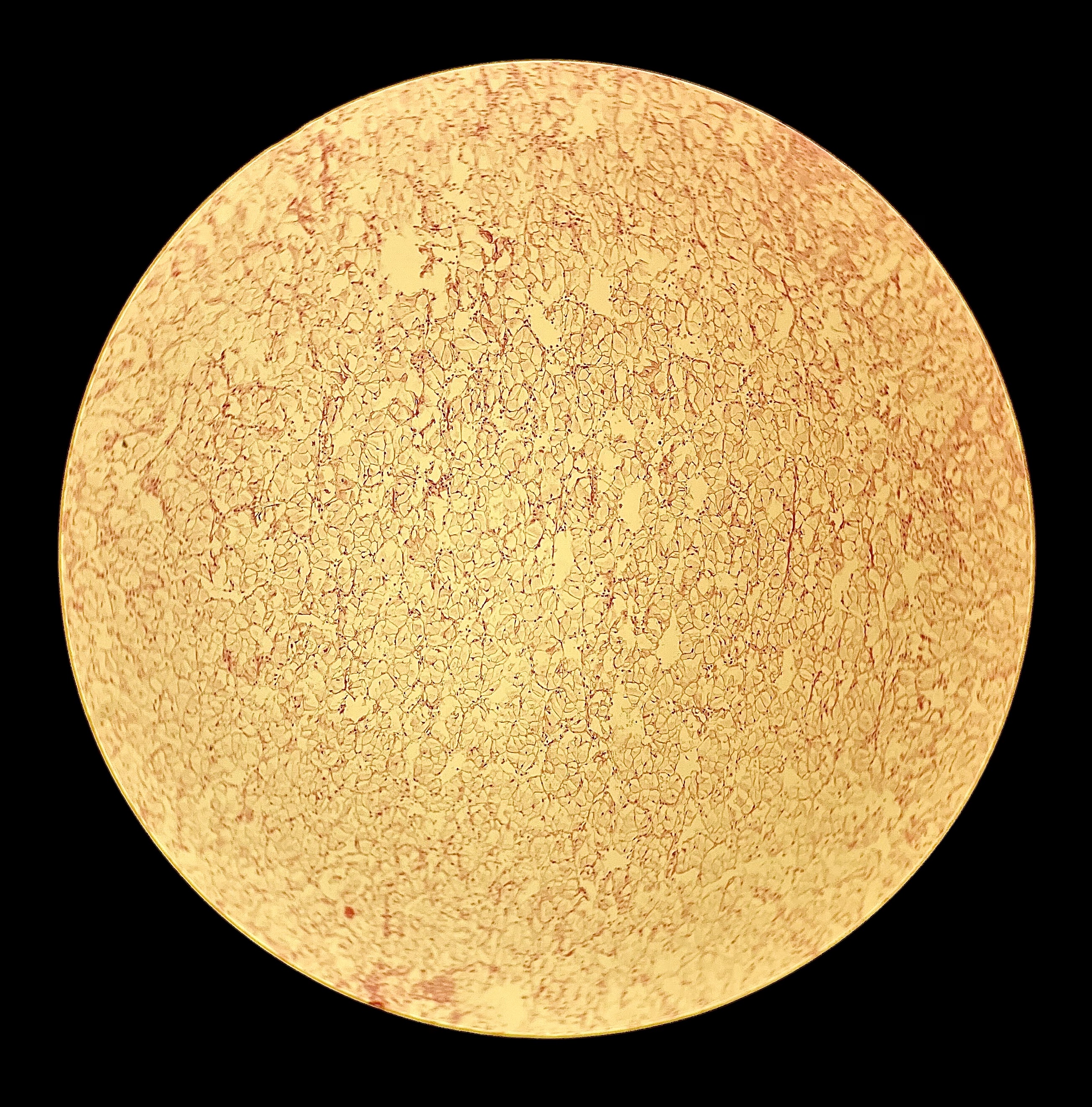
Diploid - late growth female (stage 2) (D03)
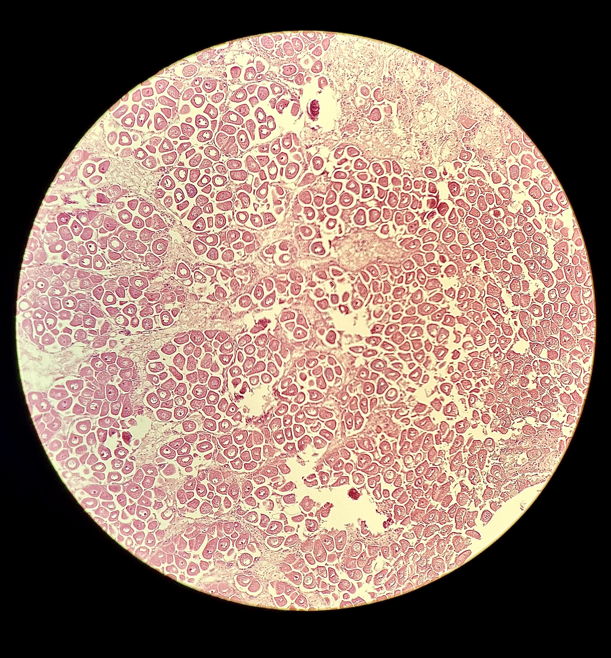
Diploid - mature female (stage 3) (D08)
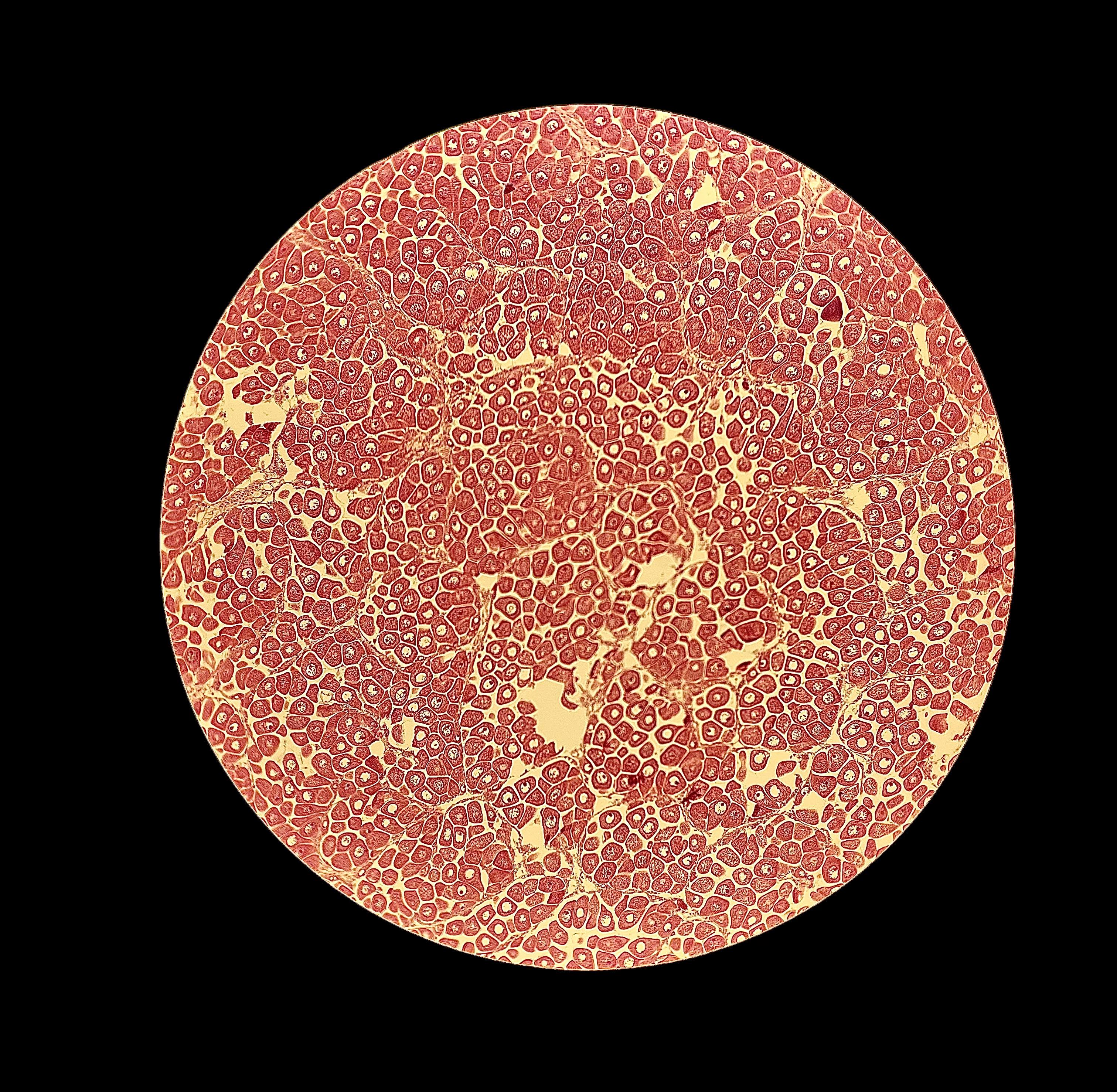
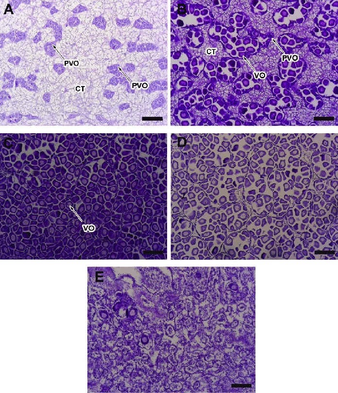
Diploid - mature female, may exhibit early spawning (stage 3-4) (D04)
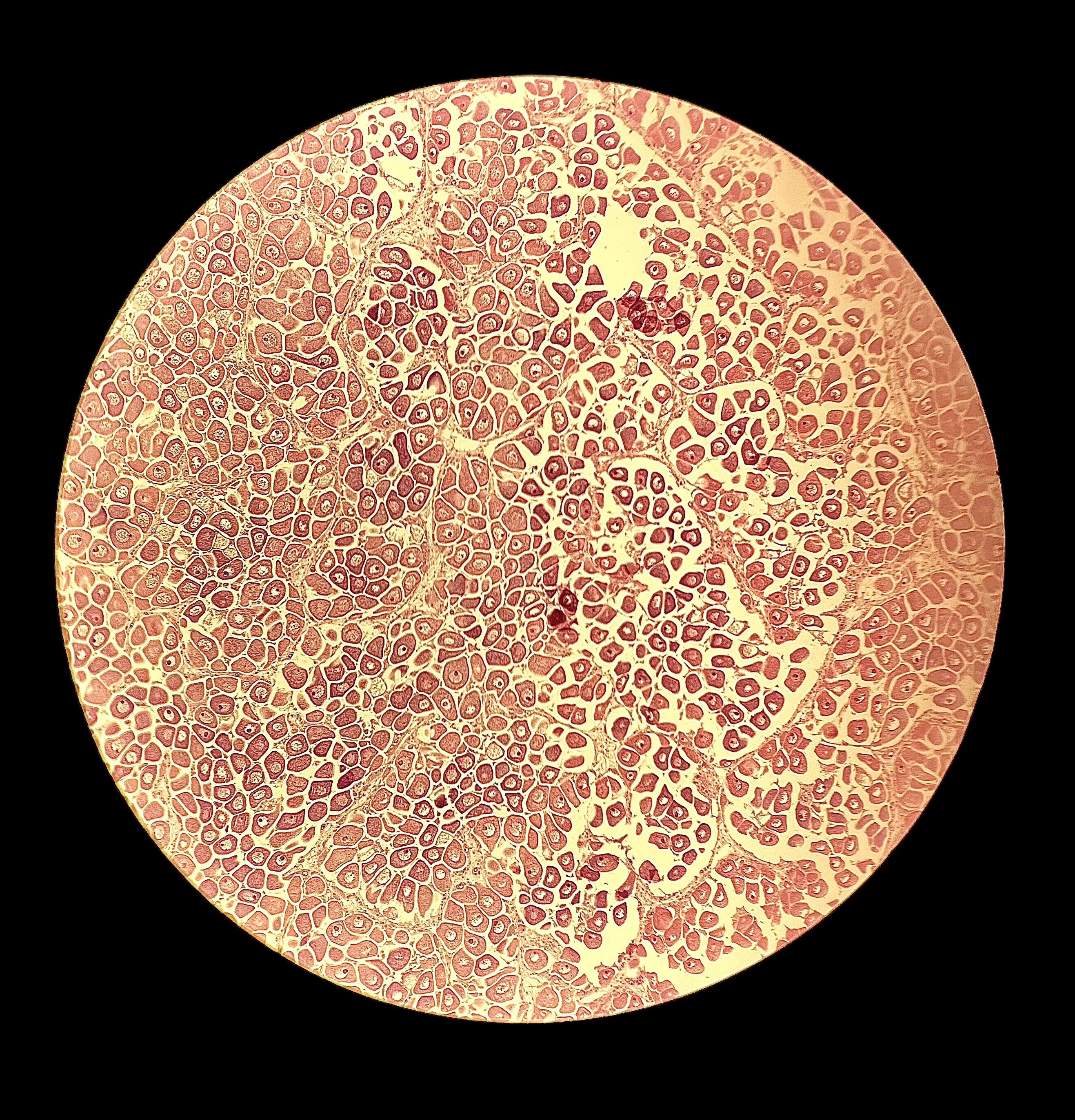
- this may be early spawning due to the open space within follicles on the right side of the image above and as seen in the image below (box C shows a mature female, and box D shows a spawning female) (Ezgata-Balic et al. 2020):
Diploid - stage could not be determined due to low presence of gonads (D02)
.jpg)
Diploid - hermaphroditic stage 3. This oyster may exhibit protogyny (rare but does occur), because there appear to be residual oocytes and a majority of male reproductive cells. Both the female and male gonads seem to be ripe, which is why this is categorized as a stage 3 hermaphrodite (D06)
.jpg)
Diploid - mature male (stage 3) (D09)
.jpg)
Triploid - resting (stage 0) (D07)
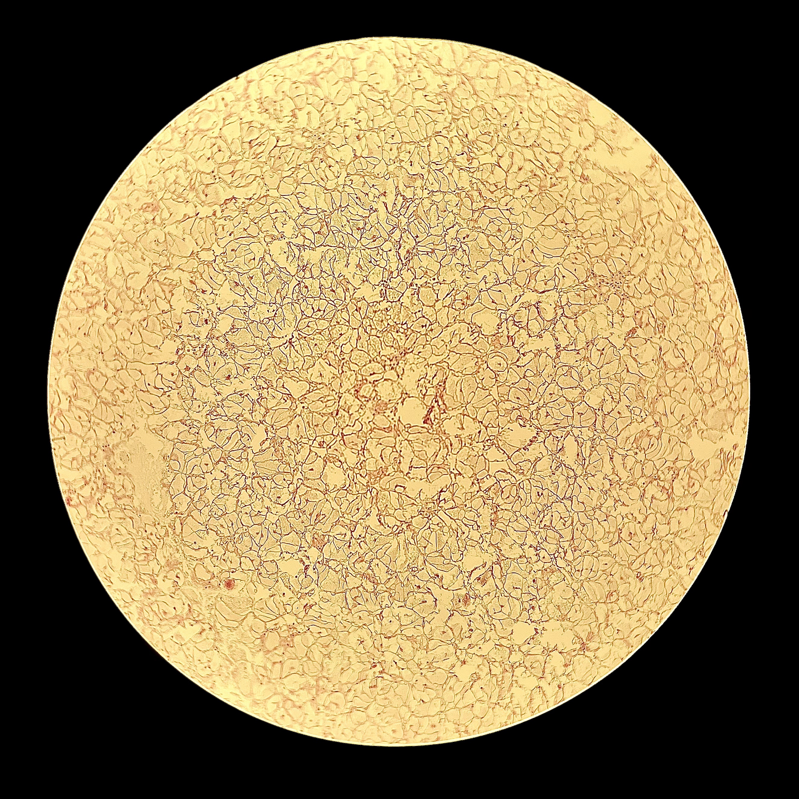
Triploid - oligo female (“follicles are lined with beta gonia and contain few to no oocytes” (Matt & Allen 2020), early growth (stage 1))
T06 10x:
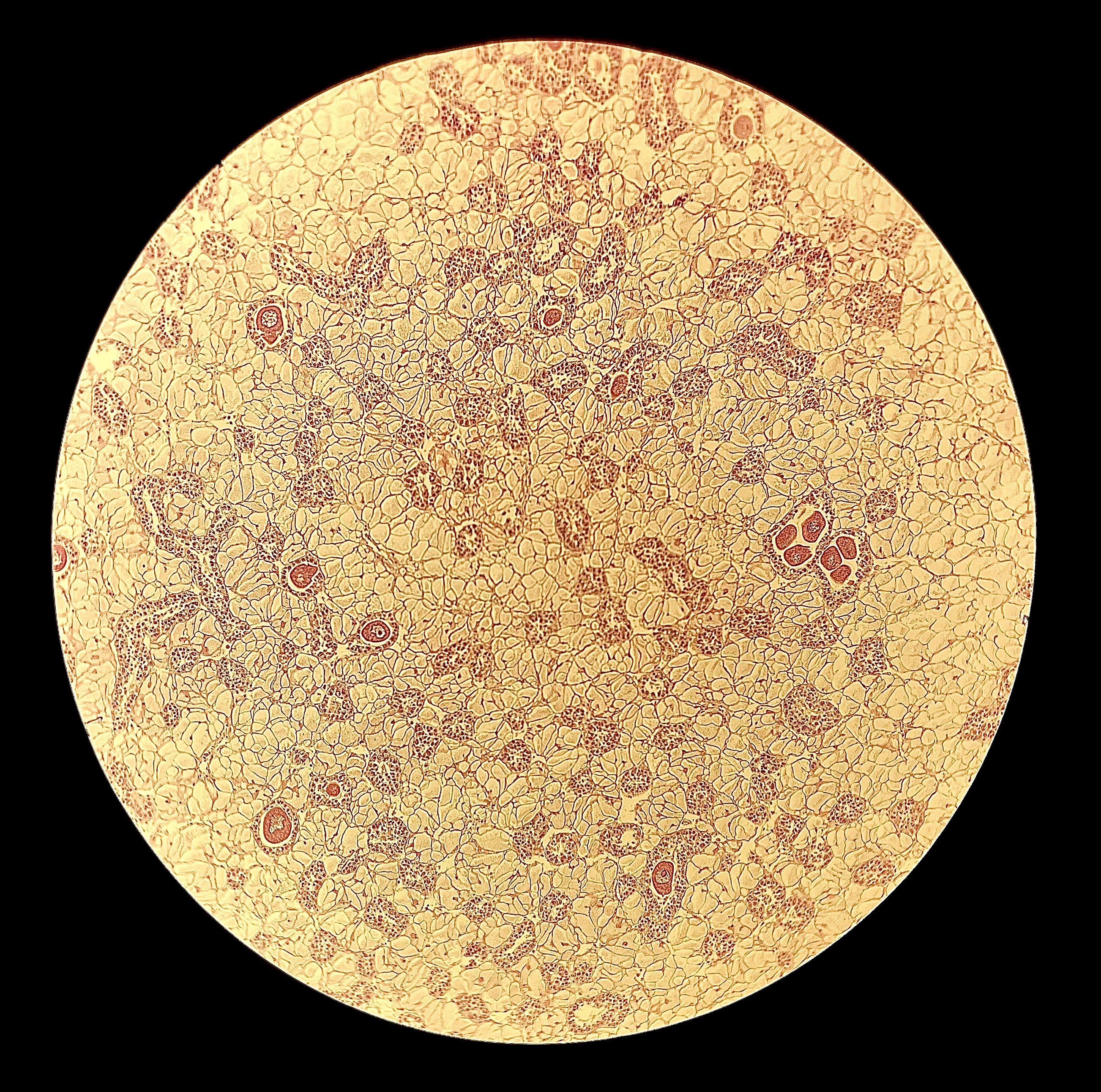
T06 40x: 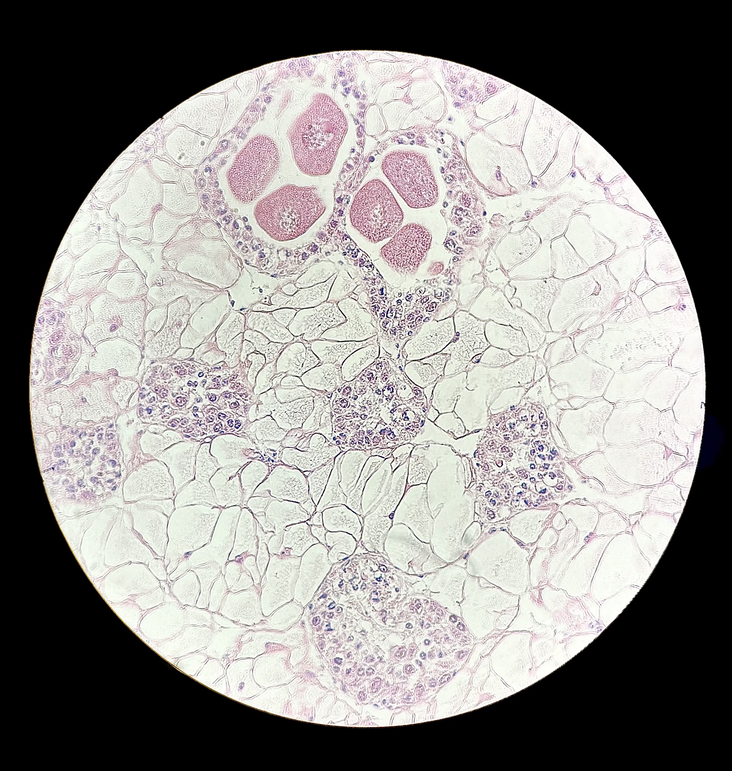
T03 10x: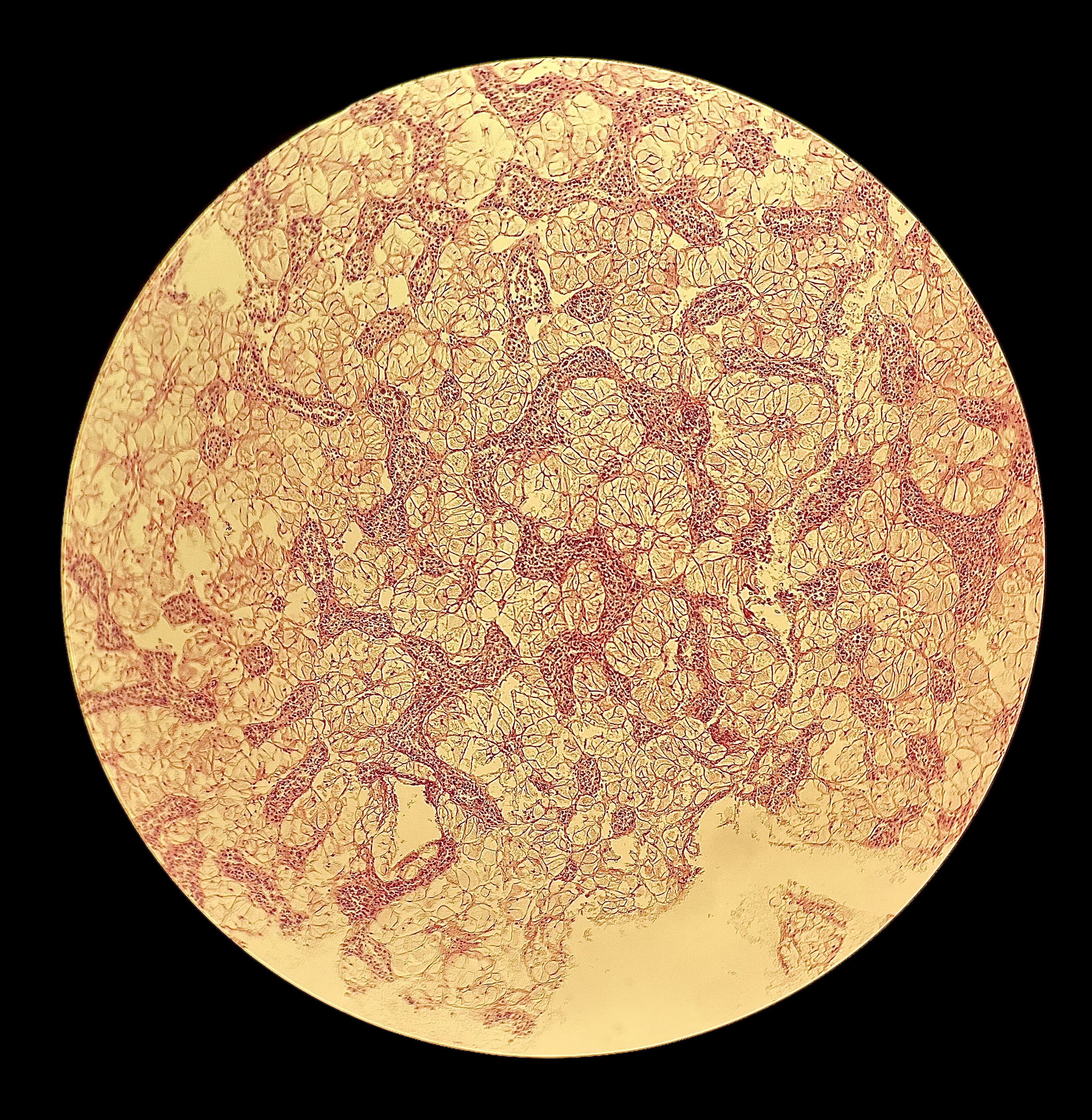
T03 40x: 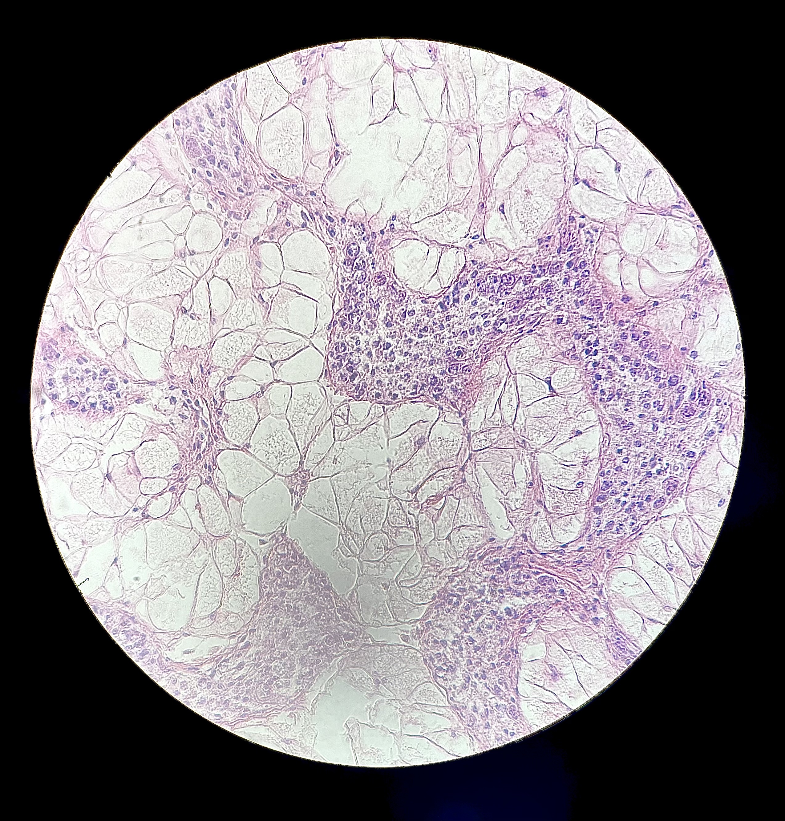
Useful papers