
Staging for Matt Part 2
I worked on staging diploid and triploid oyster samples for Matt. I took pictures of each H&E slide under a microscope under 4x, 10x, and 100x magnifications and recorded findings in this google sheet.
While staging diploids I used techniques/findings from this paper. While staging triploids I used techniques from this paper.
In a previous notebook post I included pictures of each diploid and triploid stage. In this post, I will be posting images that I was not certain about in hopes that someone will be able to help me figure those out.
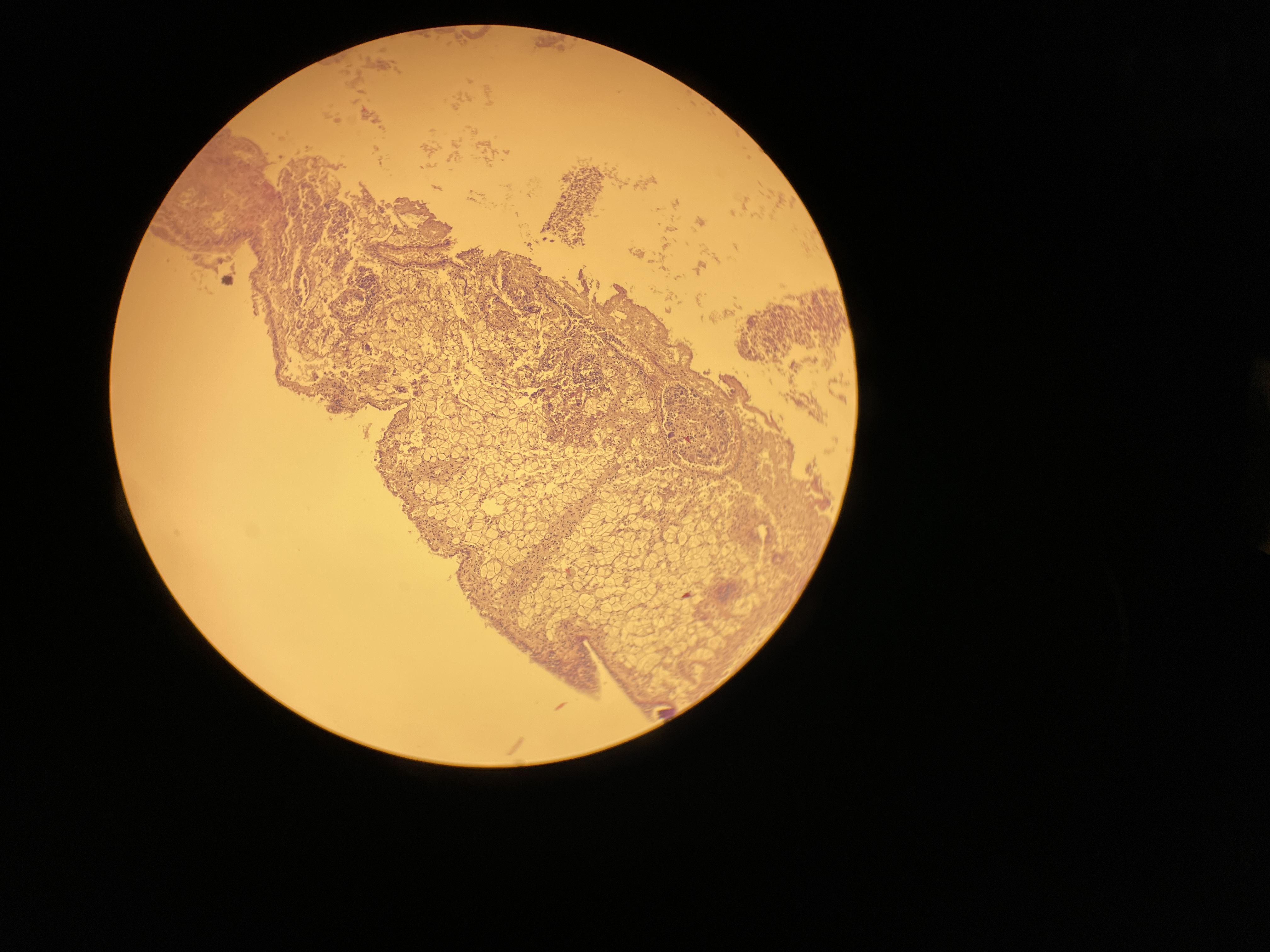
N74
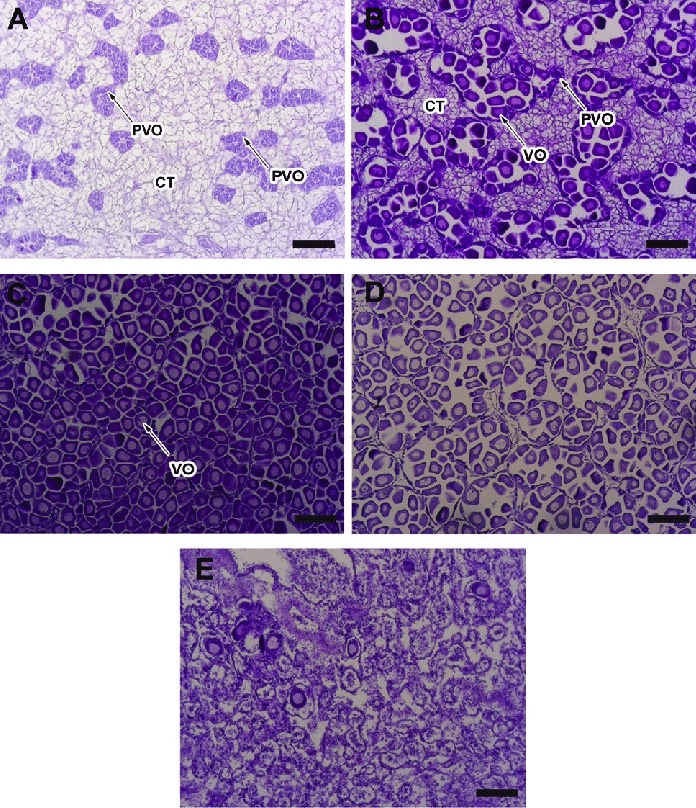
I had trouble figuring out if this individual (diploid) was spent or if the quality of the sample was just low. For reference, here is an image of a spent female from a research paper (seen in box E).
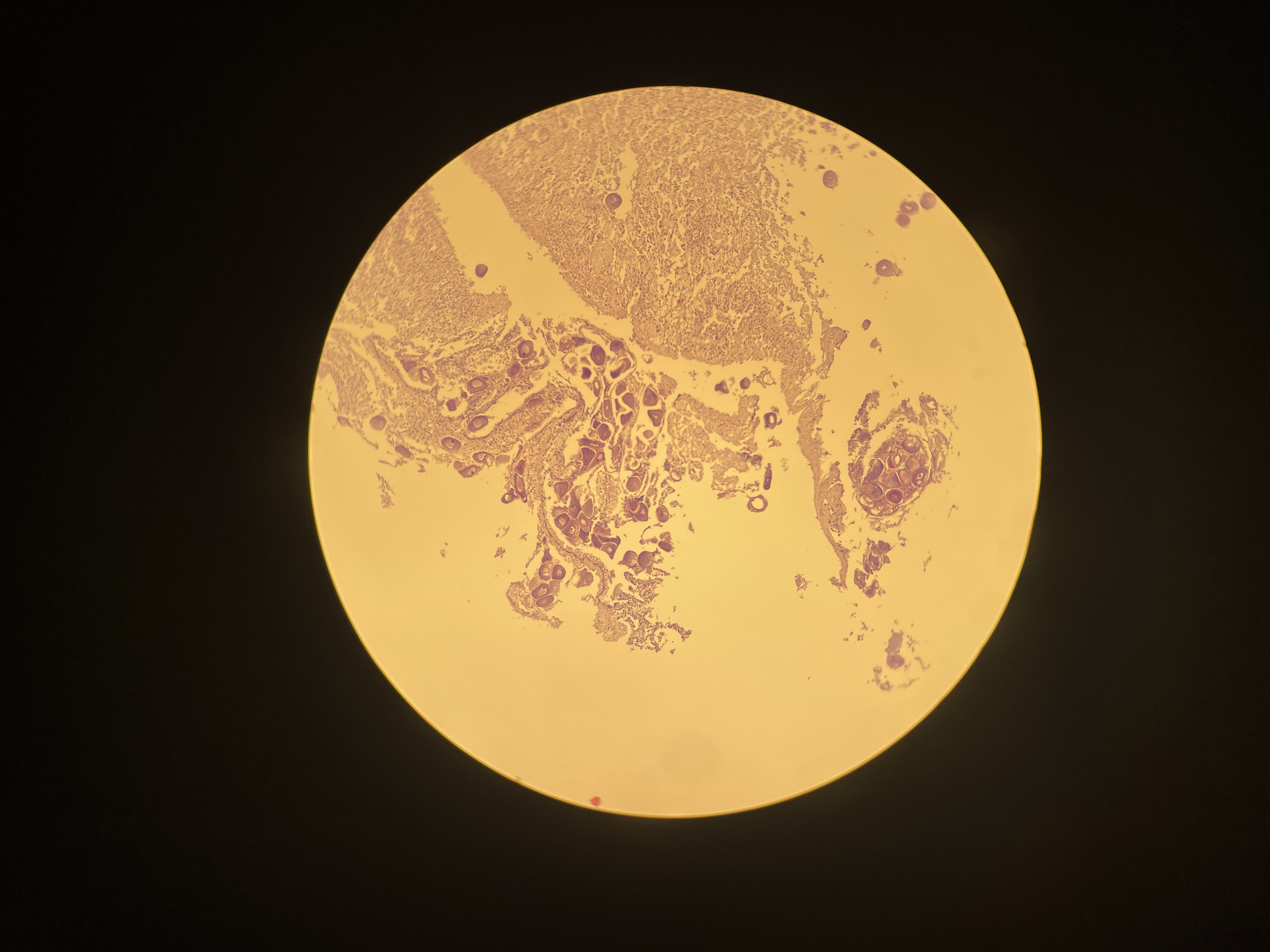
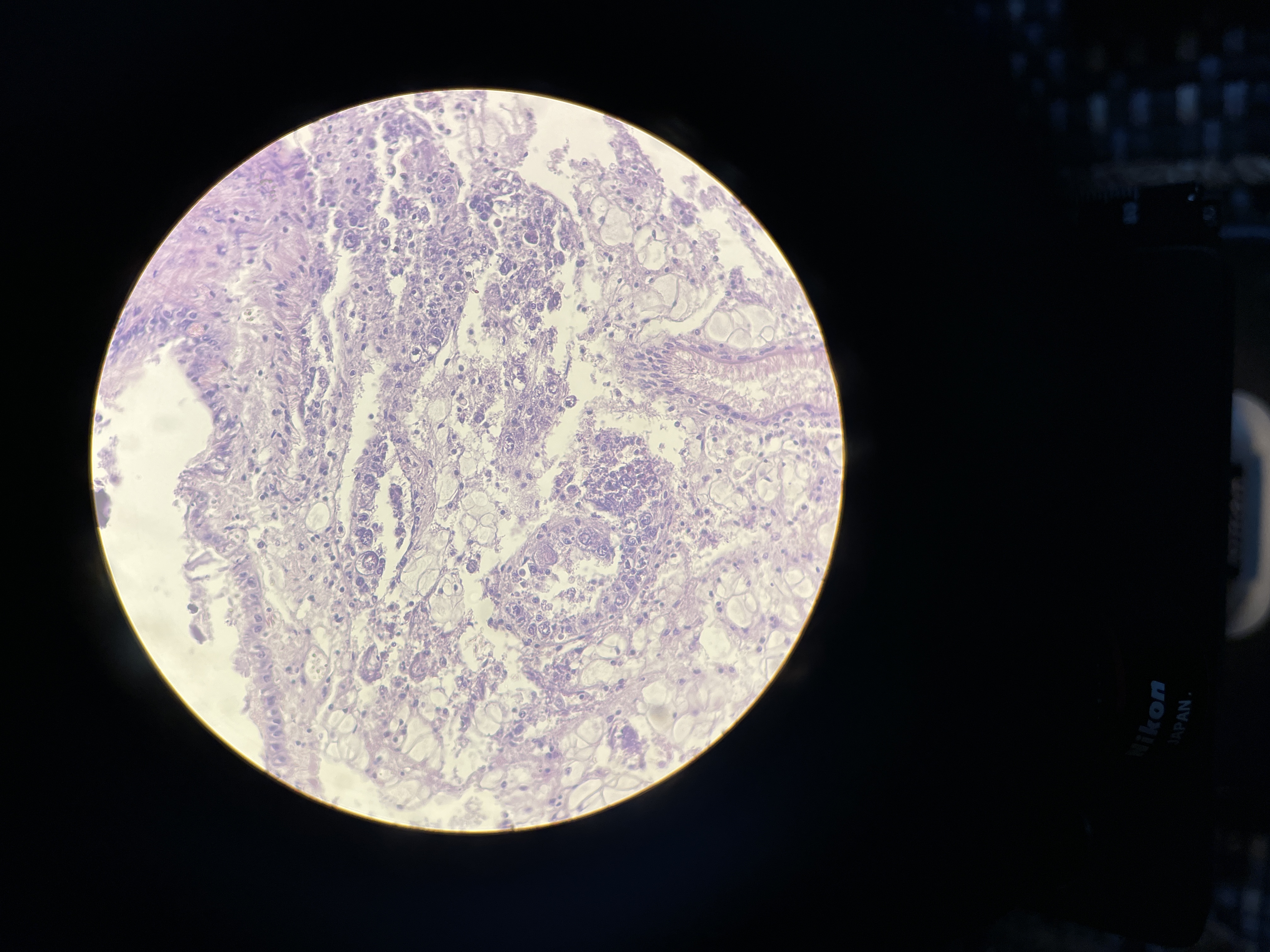
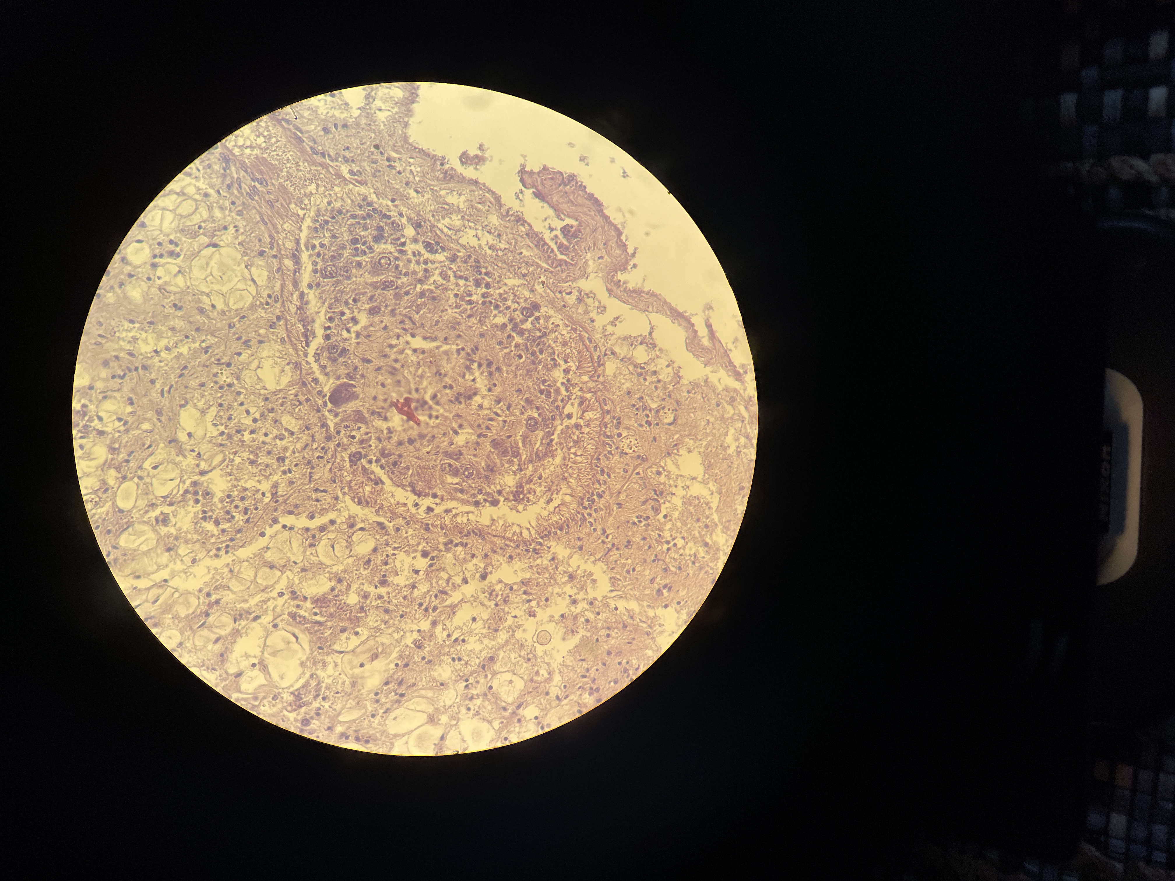
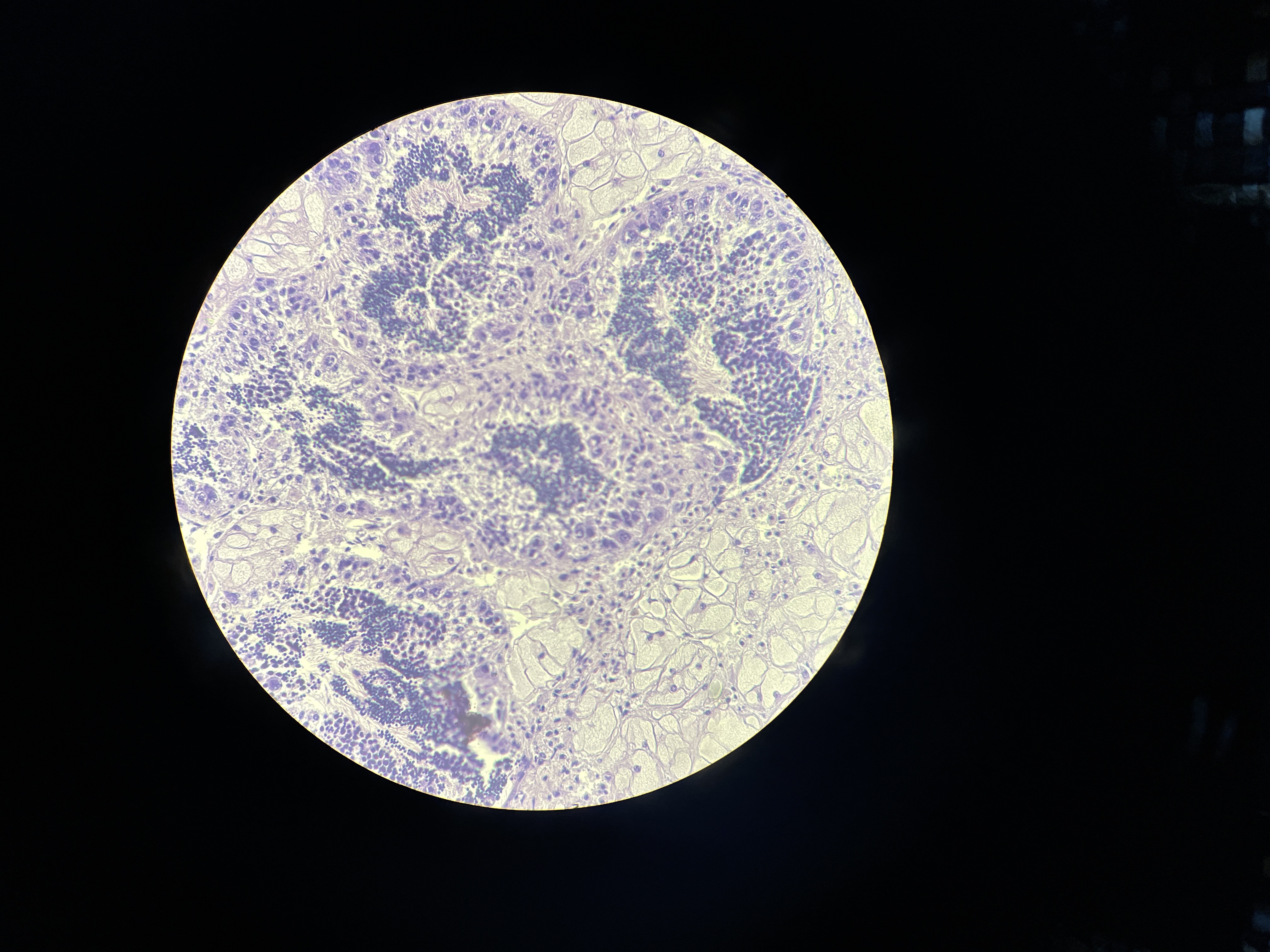
N79
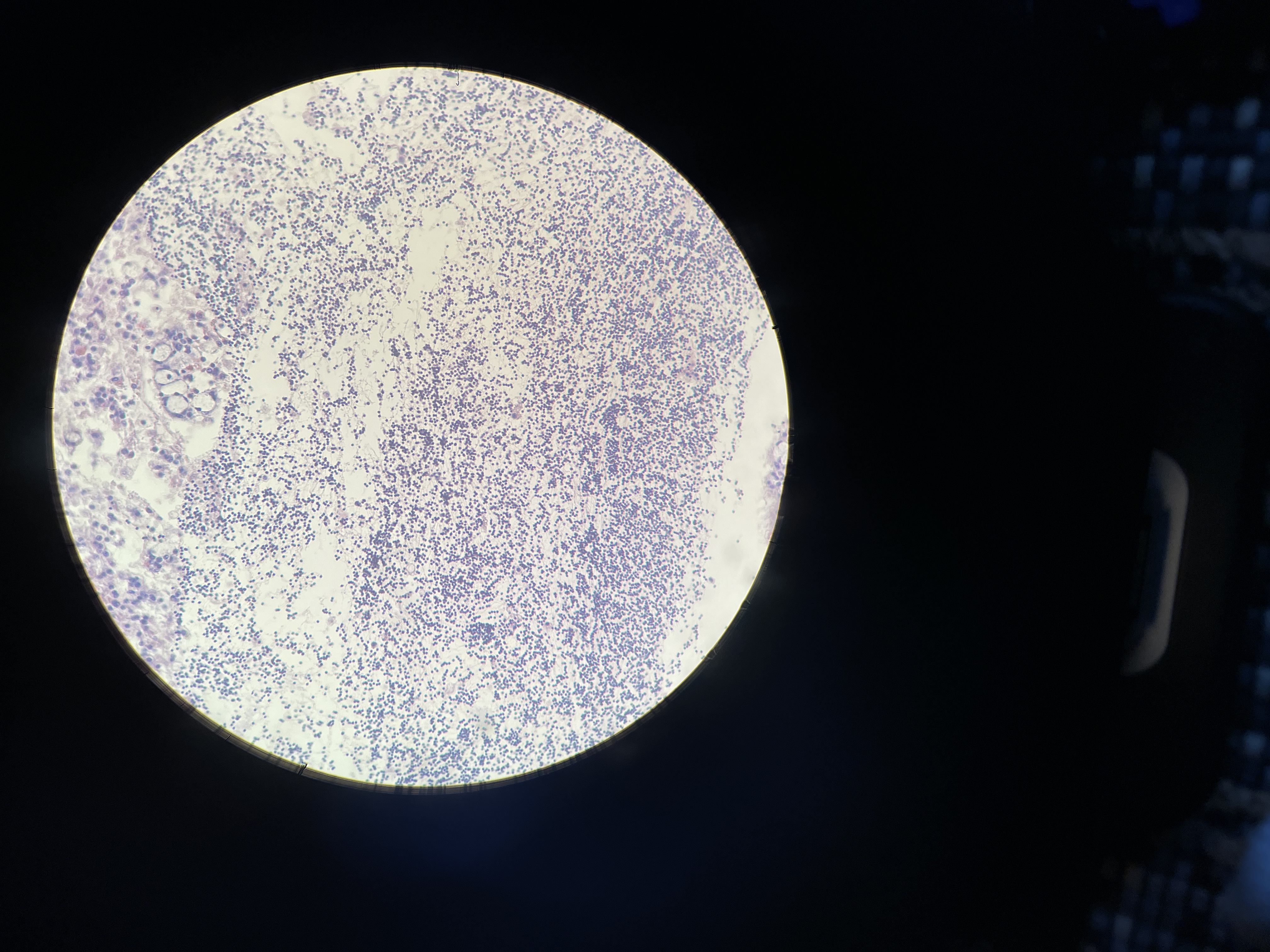 I did not know how to label this individual either. I’m just not quite sure what I am looking at with this sample. The second image seems to be primary spermatocytes, but I don’t see any follicles…
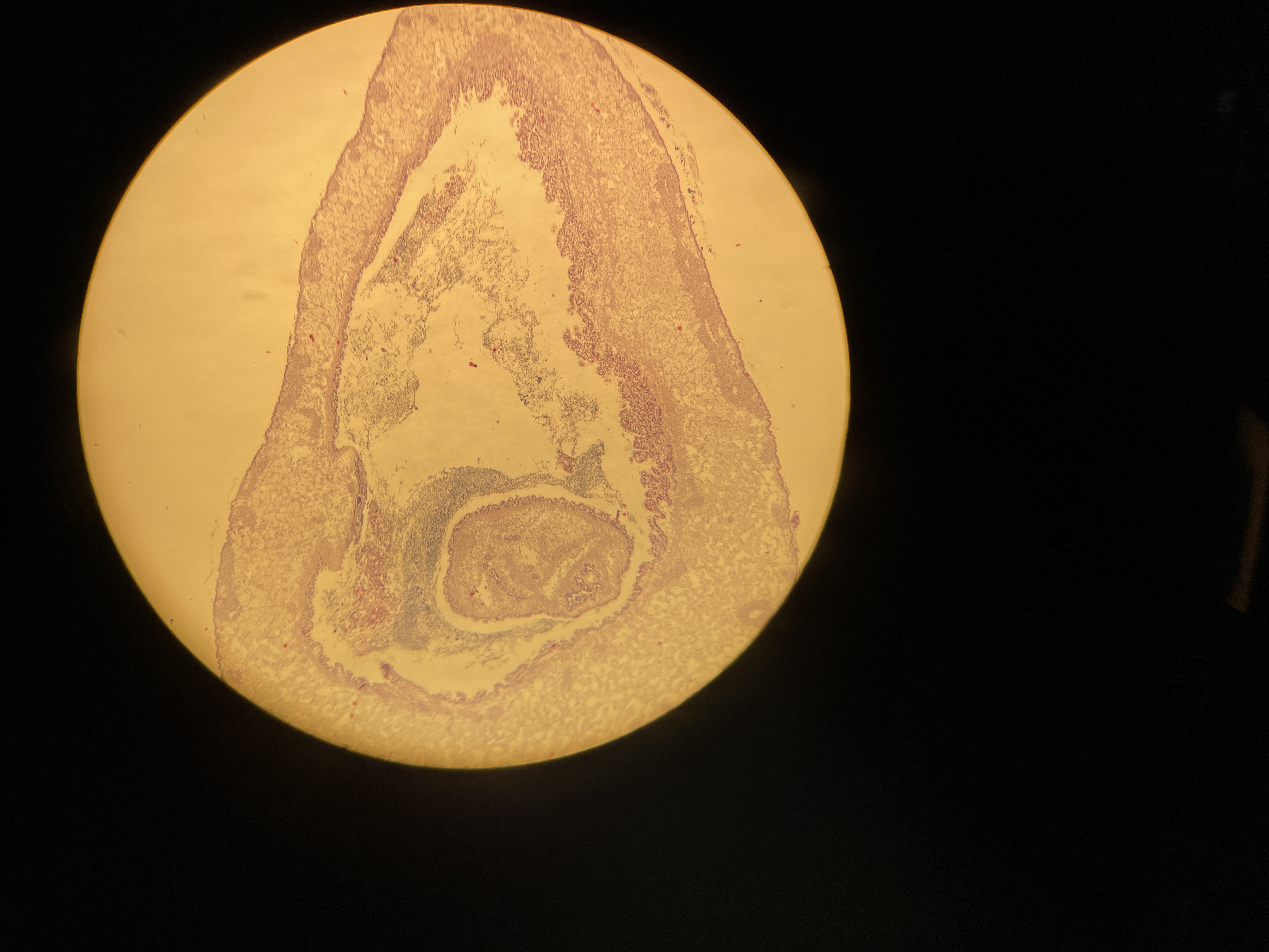
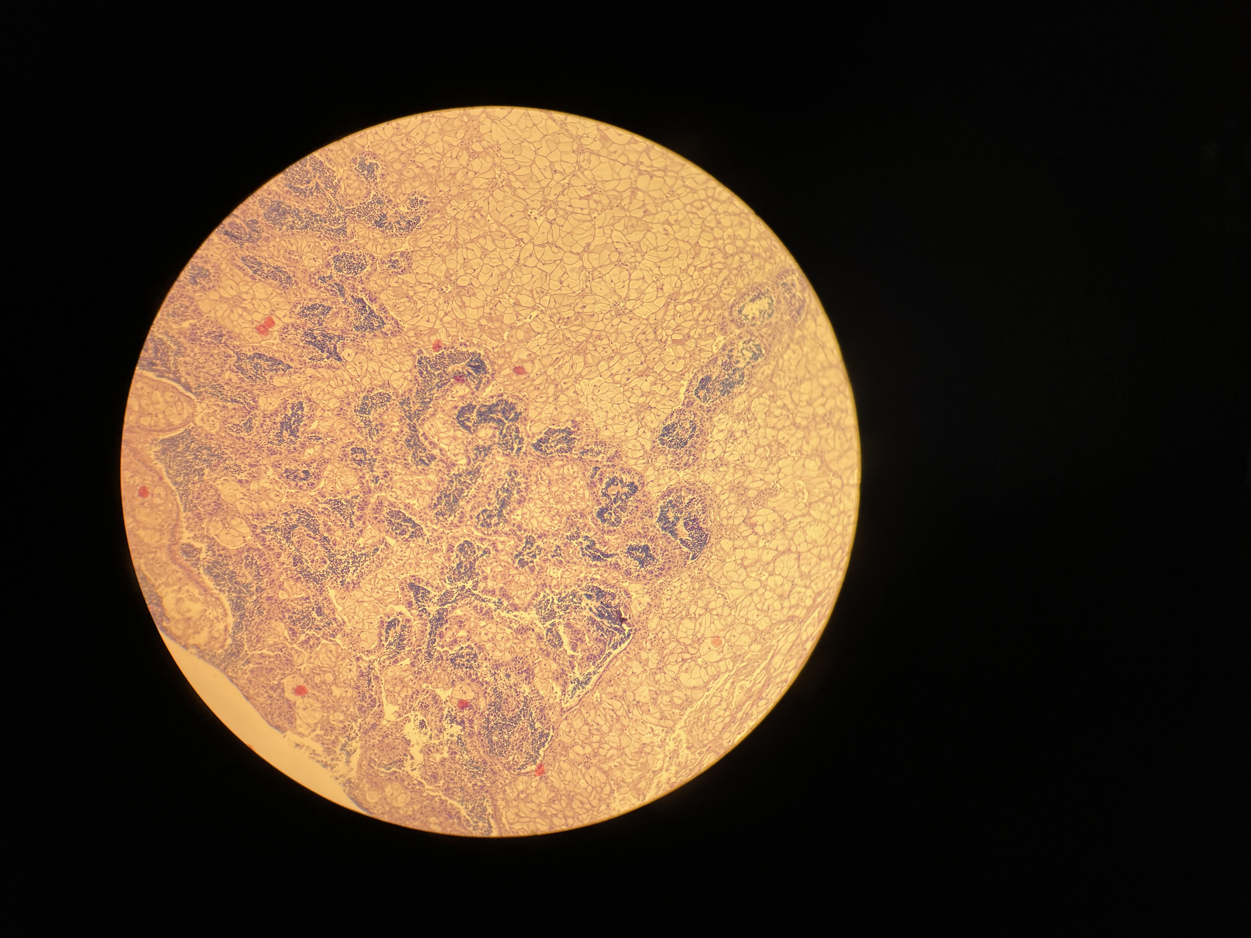
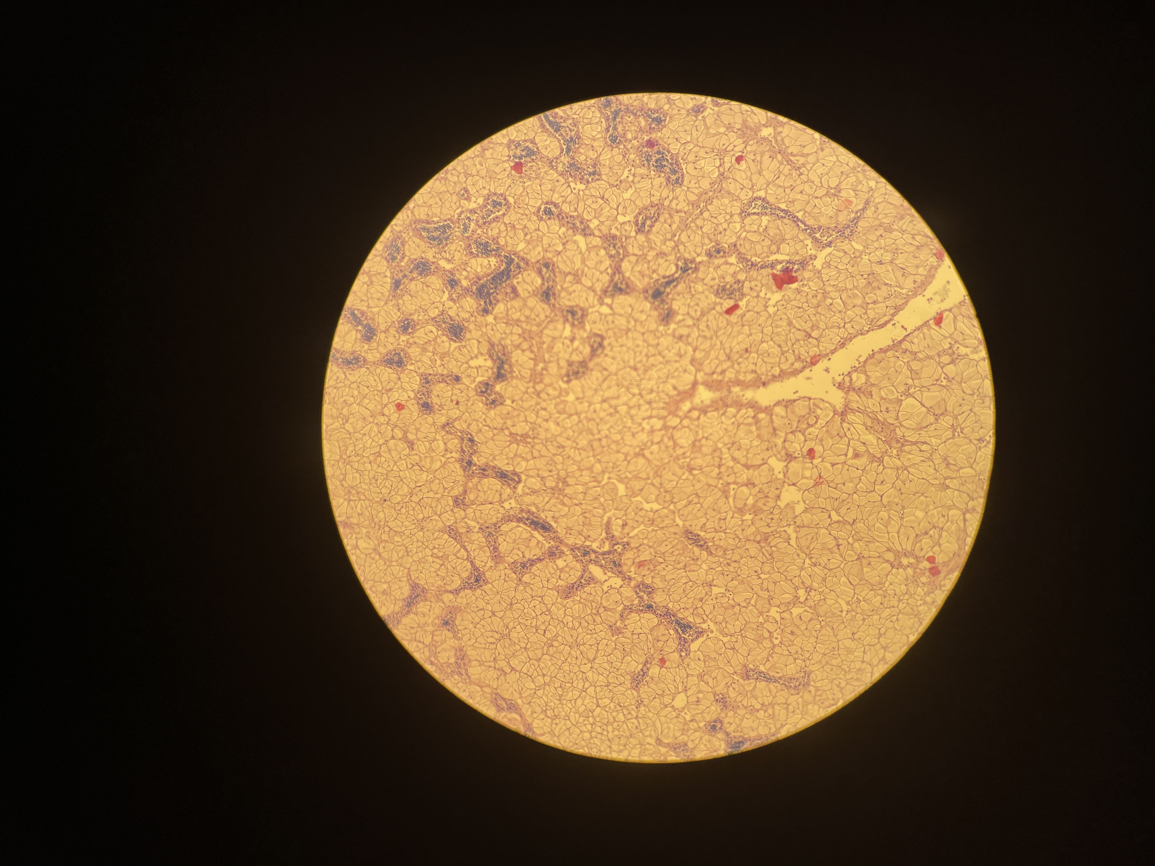
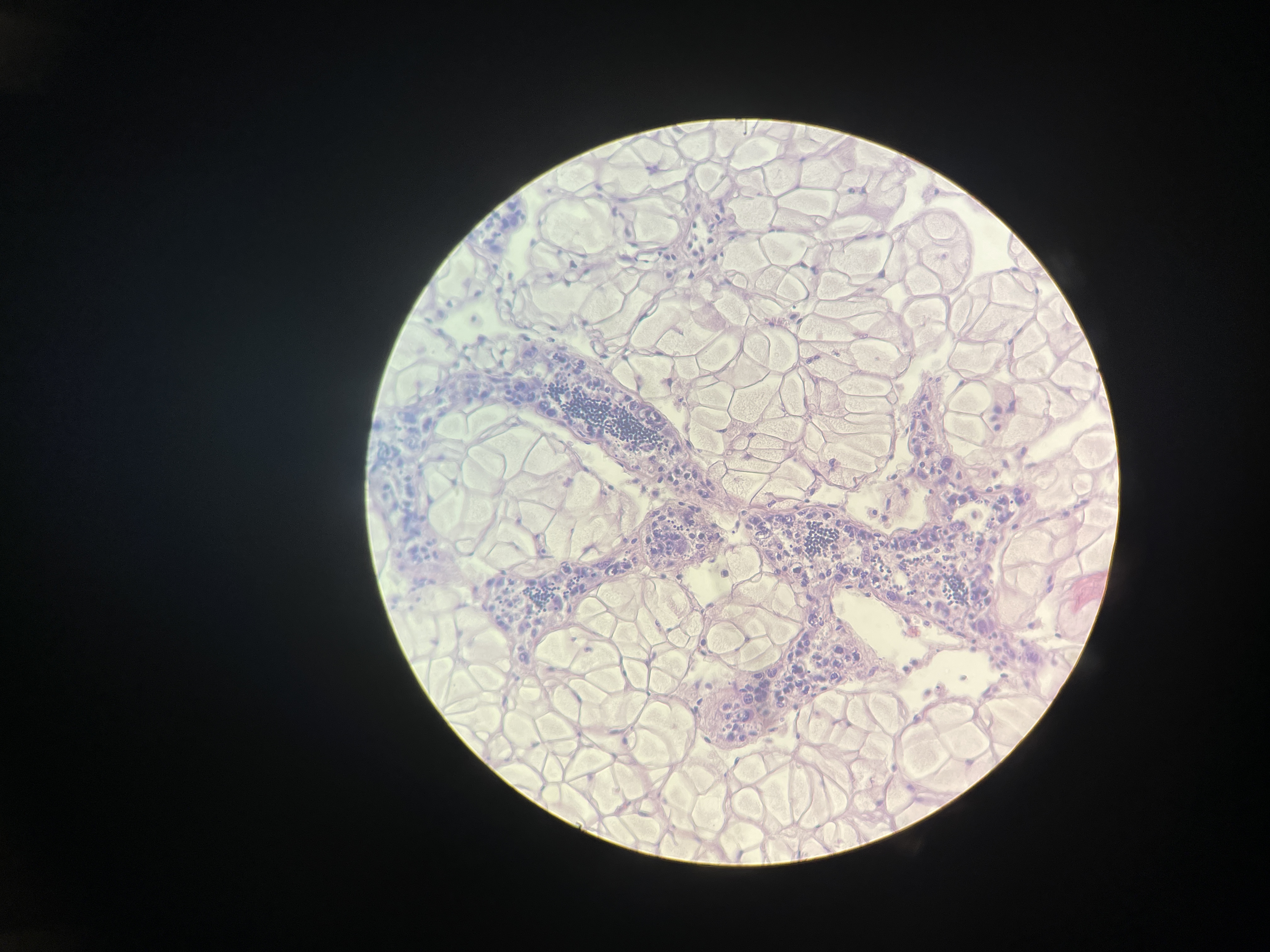
X73
 I did not know if this was gonad tissue or digestive tissue or what I was seeing here.
Virilescent females (VF)
One type of triploid that I did not observe last time was virilescent females - classified by the presence of spermatogenic cells within follicles lined with Beta gonia. Here are some images of virilescent females observed during this round of sampling:
Thanks for taking the time to read this! :)